AI-supported molecular cancer diagnosis for brain tumors
Researchers at the Hopp Children’s Cancer Center Heidelberg (KiTZ), the German Cancer Research Center (DKFZ), the Heidelberg Medical Faculty (MFHD) of Heidelberg University, and Heidelberg University Hospital (UKHD) have taken a decisive step toward more precise diagnosis of brain tumors. The latest version of the internationally used AI-based Heidelberg CNS Tumor Methylation Classifier can identify more than 180 tumor types—twice as many as the previous version. This advancement helps physicians determine tumors of the central nervous system (CNS) more accurately, enabling more targeted and less invasive treatment planning.
The Hopp Children’s Cancer Center (KiTZ) is a joint institution of the German Cancer Research Center (DKFZ), Heidelberg University Hospital (UKHD), and Heidelberg University (Uni HD).

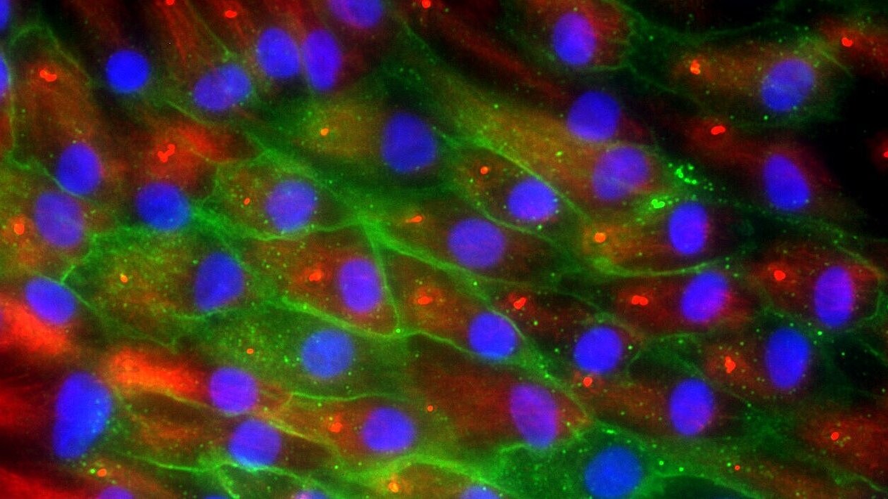
Brain tumors can be accurately diagnosed based on their methylation profiles. (C) Dominik Sturm/KiTZ
For a long time, cancer diagnosis relied solely on microscopic examination, and until recently, most brain tumors were classified mainly based on their microscopic characteristics. Meanwhile, additional molecular analyses have become a central pillar of modern diagnostics in neuro-oncology. According to the World Health Organization (WHO), such analyses are considered desirable or even indispensable for precise classification of different tumor types
The so-called Methylation Classifier is an AI-based method that evaluates tiny chemical modifications on the surface of genetic material—known as DNA methylation—to determine the origin and type of a tumor. “These epigenetic traces act like a molecular fingerprint and allow for the clear classification of tumors of the central nervous system, of which there are more than 100 subtypes,” says Felix Sahm, Professor of Neuropathology at the Medical Faculty Heidelberg of Heidelberg University, Deputy Medical Director of the Department of Neuropathology at Heidelberg University Hospital (UKHD), scientist at KiTZ, and one of the two senior authors of the study.
In the new version 12.8, the classifier was trained using approximately 7,500 tumor samples—nearly three times as many as in the previous version. As a result, the number of recognizable tumor types increased from 91 to 184. This was made possible through close collaboration with more than 100 clinics and research institutions worldwide, as well as data from an online platform where neuropathologists share their analyses.
Originally developed at the Hopp Children’s Cancer Center Heidelberg (KiTZ), the German Cancer Research Center (DKFZ), the Medical Faculty Heidelberg (MFHD) of Heidelberg University, and Heidelberg University Hospital (UKHD), the method uses a subfield of AI called machine learning to automatically analyze methylation patterns of tumor samples. It provides a probability score for each result, enabling pathologists to assess the reliability of a given diagnosis.
The clinical potential of the method became evident in the analysis of pediatric tumors across different patient cohorts: by combining molecular data with classical tissue analyses, previously misclassified cases could be corrected. Some tumors that had been classified as malignant were actually less aggressive, meaning that the affected children’s chances of survival were better than initially assumed.
“In such cases, treatment could be less intensive,” emphasizes David Jones, department head at KiTZ and DKFZ. “This means the method can help determine tumor types more precisely, improve treatment decisions, and more accurately assess the prognosis of patients with CNS tumors.”
The AI-assisted method was first published in 2018 in the renowned journal Nature and made freely available worldwide through an online platform. Since then, the Heidelberg classifier has been used by pathologists across the globe. More than 160,000 brain tumor samples from all continents have been analyzed to date.
After initially being available only for research purposes, the methylation classifier was made available worldwide in 2022 as a diagnostic tool through the spin-off Heidelberg Epignostix GmbH. In addition, an international consortium was established to make the entire process—from data generation to AI-based analysis—accessible in low-income countries worldwide.
Original publication:
Sill, M., Schrimpf, D., Patel, A., Sturm, D., et al. (2025). Advancing CNS tumor diagnostics with expanded DNA methylation-based classification. In: Cancer Cell (online publication, December 4, 2025). DOI: 10.1016/j.ccell.2025.11.002
New version of the Heidelberg methylation classifier:
https://epignostix.com/