Huntington’s Disease Biomarkers: Key Molecular Targets and Antibody Tools for HD Research
Proteins to Watch in Huntington’s Disease Research
When developing assays to study Huntington’s disease (HD), scientists have a rare advantage: they know the exact genetic cause. But that hasn’t made the biological mechanism any easier to tackle. Despite decades of research, effective treatments remain limited for this progressive degenerative disease.2 HD is inherited in an autosomal-dominant manner, passing on a pathogenic CAG repeat within exon 1 of the HTT gene, which results in production of the mutant huntingtin protein (mHTT) with an expanded polyglutamine tract.1 Over time, this mutation drives protein misfolding, aggregation, and neuronal dysfunction, and the brain’s striatum bears the brunt of the damage as medium spiny neurons (MSNs) are progressively lost.1,2
Because HD follows autosomal dominant inheritance, the mutation is detectable decades before symptoms appear, giving clinical researchers a rare opportunity to study neurodegeneration at its earliest, most treatable stages.2 This head start means scientists can develop assays to examine relevant biomarkers in a genetically defined population and assess the impact of emerging therapeutics, like treatments that lower mHTT.2
Making the most of that opportunity hinges on choosing the right antibody biomarkers for your preclinical compound screens. Below, we’ve outlined key molecular targets in HD research—from the huntingtin protein itself to downstream markers of striatal dysfunction, neuronal injury, and metabolic failure—making it easier to select the right panels of validated Cell Signaling Technology (CST) antibodies for the most informative preclinical readouts.
How Huntingtin Disrupts Protein Interactions
Protein accumulation is the defining molecular feature of HD. As mHTT misfolds and aggregates, it disrupts proteins that normally regulate neuronal signaling, intracellular trafficking, and synaptic organization.1,2 Antibodies that target huntingtin and its interacting partners are essential tools for studying the pathobiology of HD—and for evaluating the growing number of HTT-lowering therapeutic strategies in development.
CST antibodies are highly specific, rigorously tested, and available in carrier-free formats—which are free from BSA and azide—making them ideal tools for your ELISA and sandwich assays. Contact us to inquire about carrier-free antibodies.
#1. Huntingtin (HTT / mHTT)
Huntingtin is a large (~350 kDa) cytosolic protein expressed throughout the body, with the highest expression in the brain—particularly in the striatum, neocortex, hippocampal formation, and cerebellar cortex.1In HD, the expanded polyglutamine tract in mHTT promotes misfolding and proteolytic cleavage, leading to aggregation-prone N-terminal mHTT fragments, which subsequently impact transcription, axonal transport, mitochondrial homeostasis, and autophagy.1,2
As HTT-lowering strategies continue to be investigated, measuring mHTT in CSF and plasma has become a key pharmacodynamic endpoint—one that requires antibodies with proven sensitivity and specificity for accurate readouts in bead-based immunoassays and ELISAs.2 A 2024 consensus platform study comparing 20 commercial huntingtin antibodies found significant variability in performance across reagents, underscoring the importance of using rigorously validated tools for any assay-based mHTT detection workflow.3 Antibodies developed at CST undergo unparalleled, application-specific validation using a multilayered strategy before they reach the market—so researchers can trust that our antibody tools will consistently deliver specific, reproducible results in their recommended applications.
CST offers a portfolio of highly validated recombinant monoclonal antibodies that target distinct epitopes across the full-length huntingtin protein, from the N-terminal region near the polyQ domain through the N-HEAT repeats, bridge domain, and C-HEAT repeats. They enable detection across a range of disease models—including human or mouse tissues and primary cells, such as iPSC-derived neurons—and research applications, including ELISAs, high-content screening (HCS), and immunofluorescence (IF).
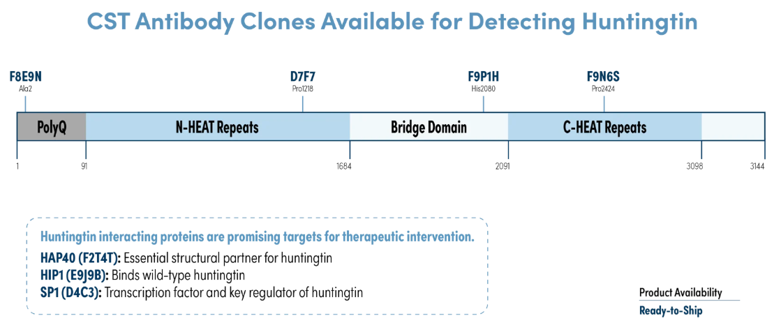
- Huntingtin (D7F7) Rabbit Monoclonal Antibody (Biotinylated) #93535 – ELISA+; H, M, R
- Huntingtin (D7F7) Rabbit mAb (BSA and Azide Free) #31873 — WB, IF; H, M, R
- Huntingtin (F8E9N) Rabbit mAb #73232 — WB, IHC, IF; H, M, R
- Huntingtin (F9P1H) Mouse mAb #58445 — WB, IF; H, M, R