MAGIC: AI-assisted laser tag illuminates cancer origins
EMBL researchers have developed a new AI tool, which, through a game of molecular laser tag, identifies cells that can shed light on the earliest origins of cancer
Summary
- During cell division, chromosomal abnormalities – defects in chromosome number or structure – can occur, which can cause particularly aggressive forms of cancer.
- A new study has shed light on how chromosomal abnormalities arise in normal cells, the rates at which they do so, and how these rates are affected by various factors.
- To do this, the scientists developed a novel and versatile artificial intelligence (AI) tool called MAGIC, which combines automated microscopy and image analysis with single-cell genome sequencing.
- Understanding the molecular origins of cancer in this way could pave the way for better genetic detection tools and potential medical prevention strategies in the future.
The human body relies on precise genetic instructions to function, and cancer begins when these instructions get scrambled. When cells accumulate genetic errors over time, they can break free of the normal controls on their growth and divide excessively. Chromosomal abnormalities – numerical and structural defects in chromosomes – are a common first step in this process, often contributing to normal cells turning cancerous.
A new AI tool developed by researchers in the Korbel Group at EMBL Heidelberg now offers a powerful method to gain deep insights into how such chromosomal abnormalities arise in the first place. This knowledge could eventually help scientists understand the origin of cancer.
“Chromosomal abnormalities are a main driver for particularly aggressive cancers, and they’re highly linked to patient death, metastasis, recurrence, chemotherapy resistance, and fast tumour onset,” said Jan Korbel, senior scientist at EMBL and senior author of the new paper, published in the journal Nature. “We wanted to understand what determines the likelihood that cells undergo such chromosomal alterations, and what’s the rate at which such abnormalities arise when a still normal cell divides.”
The idea of chromosomal abnormalities leading to cancer is not new. In fact, more than a century ago, German scientist Theodor Boveri was the first to hypothesise, based on microscopy studies, that abnormal chromosomal contents in a cell contribute to the development of cancer.
However, since only a small fraction of cells display chromosomal abnormalities at a given time, and these cells often die (or are killed off) via a natural selection process, their detection has previously proved to be a key challenge. Scientists had to manually spot such cells under the microscope, and only a handful of cells could be isolated at a time for further analysis.
Marco Cosenza, Research Scientist in the Korbel Group, hit upon the solution to this problem after working with other teams at EMBL wrestling with similar challenges. He and his collaborators developed a new, autonomous system that combines automated microscopy, single-cell sequencing, and AI, which they named machine learning-assisted genomics and imaging convergence – or MAGIC.
‘Laser tag’ to precisely identify and mark cells
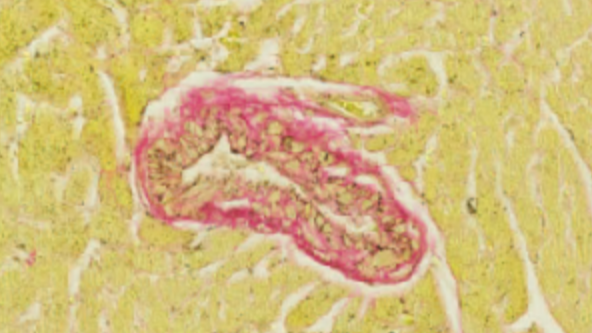
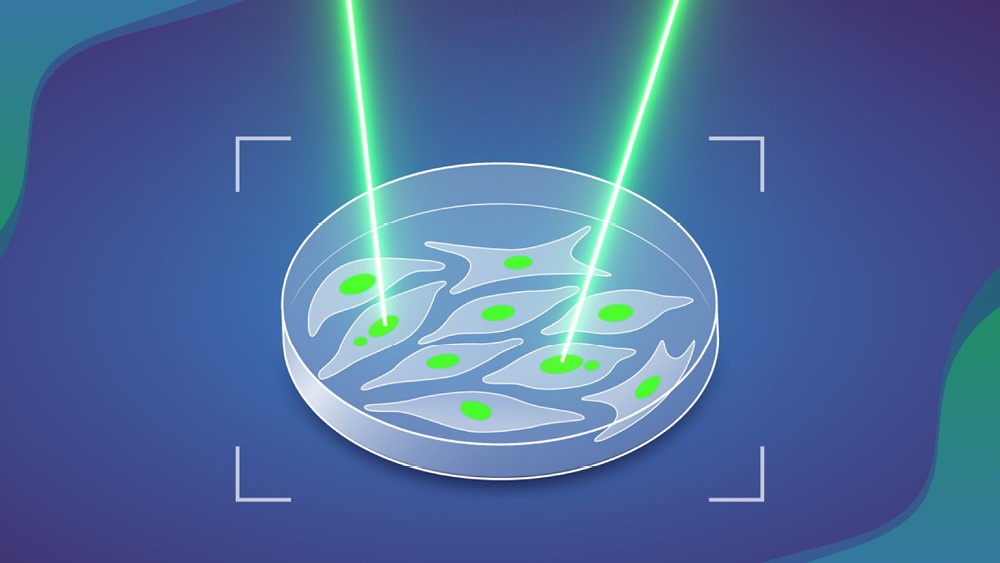
Essentially, MAGIC operates like a fully automated game of laser tag. It spots ‘enemies’, or cells, with a particular kind of visible feature. For this study, the scientists focused on a cellular structure called a ‘micronucleus’. Micronuclei are tiny enclosed compartments inside cells that contain a small portion of the cells’ DNA, broken off from the bulk of the genome. Cells with micronuclei tend to produce new chromosomal abnormalities, which makes them more likely to turn cancerous.
Once cells with micronuclei are detected, the system ‘tags’ them using a laser. For this, the scientists used a photoconvertible dye – a fluorescent molecule that undergoes a chemical transformation if light is shone on it, changing the colour of light it emits.
“This project combined a lot of my interests in one,” said Cosenza. “It involves genomics, microscopic imaging, and robotic automation. During the COVID-19-related lockdown in 2020, I could really spend some time on learning and applying AI computer vision technologies to the biological image data we had collected before. Afterwards, we designed experiments to validate it and take it further.”
In practice, MAGIC works like this. First, an automated microscope captures a series of images of a cell sample. Next, a machine learning algorithm, trained on manually annotated datasets of micronuclei-containing cells, scans the images. When the algorithm spots cells with micronuclei, it shares their location with the microscope and instructs it to shine light specifically on those cells, permanently tagging them. The tagged cells can then easily be separated from these still-living cells using methods like flow cytometry, and subsequently be subjected to deeper analysis, e.g. by looking at their cellular genomes.
By automating the previously labour-intensive, time-consuming, and error-prone process of detecting cells with micronuclei, MAGIC allows scientists to study such cells at a scale and speed previously unheard of. In less than a day, scientists can analyse nearly 100,000 cells using this method.
The team used MAGIC to analyse chromosomal abnormalities in cultured cells originally derived from normal human cells. Their results showed that a little more than 10% of all cell divisions result in spontaneous chromosomal abnormalities of some kind and that this rate nearly doubles when a particular gene – p53, a well-known tumour suppressor – is mutated. The scientists also studied other triggers and contributors to chromosomal abnormality formation, such as the presence and location of double-stranded DNA breaks within a chromosome.
The study involved collaborations across and outside EMBL, with key contributions from the Advanced Light Microscopy Facility (ALMF) and the Pepperkok Team at EMBL Heidelberg, Isidro Cortes-Ciriano’s group at EMBL-EBI, and Andreas Kulozik’s team at the German Cancer Research Centre (DKFZ), which also forms part of the Molecular Medicine Partnership Unit (MMPU) between EMBL and the University of Heidelberg.
MAGIC is a highly versatile and adaptable technique. While the scientists trained it for this study to spot cells that had micronuclei, the algorithm can, in theory, be trained on many different kinds of datasets to detect different cellular features.
“As long as you have a feature that can be discriminated visually from a ‘regular’ cell, you can – thanks to AI – train the system to detect it,” said Korbel, “Our system therefore has potential to advance future discoveries in numerous areas of biology.”
Source article(s)
Origins of chromosome instability unveiled by coupled imaging and genomics
Cosenza M.R. et al
Nature 29 October 2025
10.1038/s41586-025-09632-5