New regulatory protein of the neuronal cytoskeleton identified

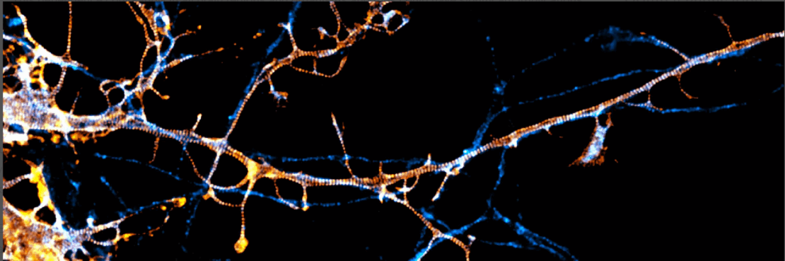
The growth and function of nerve cells is based on a lattice-like structure called membrane-associated periodic skeleton (MPS). However, how the organization of the MPS is controlled has not been elucidated until now. A team from the Max Planck Institutes for Medical Research and for Multidisciplinary Sciences has now discovered that it is regulated by the concentration of the protein paralemmin-1.
- In their study published in Science Advances, the researchers show for the first time that the protein paralemmin-1 plays a critical role as a powerful regulator in the organization of the MPS in nerve cells.
- This dependence could apply in all brain regions and organisms and thus could have significant implications for understanding MPS regulation in the human brain.
- Paralemmin-1 also plays a key role in the formation of nerve cell processes and cell growth.
© Victor Macarrón-Palacios / MPIMR
Key role of paralemmin-1 identified
Nature has developed a unique structure as a scaffold for almost all nerve cells: the membrane-associated periodic skeleton, MPS. This specialized cytoskeletal structure is located below the cell membrane and consists of numerous proteins in a periodic arrangement. The MPS is involved in various cellular processes, such as inter- and intracellular signaling. To date, many proteins have been identified that interact with the MPS, but the mechanisms underlying its organization are not yet fullyunderstood.
“Our work started with the question of whether paralemmin-1 is associated with the MPS in neurons,” explains Victor Macarrón-Palacios of the Max Planck Institute (MPI) for Medical Research in Heidelberg. “Ultimately, we were able to show that paralemmin-1 is indeed associated and that it also plays a key role: It regulates the organization of the MPS.”
Nanoscopy provide crucial insights
Visualizing the MPS is not possible with conventional microscopy techniques, only with nanoscopy. For their study, the scientists at the MPI for Medical Research used state-of-the-art fluorescence nanoscopy: the STED- and MINFLUX microscopy for which Max Planck director Stefan Hell was awarded the Nobel Prize in Chemistry in 2014.
Acollaboration between the MPI for Medical Research and the MPI for Multidisciplinary Sciences in Göttingen led to this successful project. Manfred W. Kilimann, retired university professor and guest researcher at the MPI for Multidisciplinary Sciences, initiated the study and provided the team in Heidelberg led by Elisa D’Este with the results and materials compiled in his laboratory. This enabled Victor Macarrón-Palacios to investigate the neural cytoskeleton in his doctoral thesis. These discoveries were further complemented by biochemical techniques and the expertise and support of the entire scientific team: a perfect example of interdisciplinary collaboration and synergy between the two institutes. Further contributions to this research work came from scientists at the universities of Heidelberg and Uppsala (Sweden).
© Victor Macarrón-Palacios / MPIMR
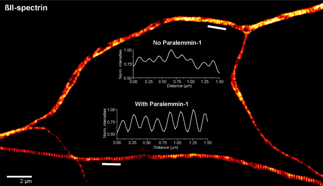
The researchers were able to show that the concentration of paralemmin-1 is of fundamental importance for controlling the nanoscale organization of the periodic scaffold of the cytoskeleton: “High levels of paralemmin-1 result in an extremely tight periodic arrangement, while lower levels lead to a poorly organized MPS,” reports Manfred W. Kilimann. Moreover, the absence of paralemmin-1 affects the electrophysiological properties of the neurons, specifically the electrical signal transmission between them. The team was also able to show that paralemmin-1’s ability to fulfill its function depends on a single tryptophan amino acid, W54.
A deeper understanding of the paralemmin protein family and MPS
“By studying paralemmin-1, we have identified a mechanism that regulates the fine structure of the MPS in neurons,” explains Victor Macarrón-Palacios. In this process, paralemmin-1 binds the protein ßII-spectrin, a major component of the neuronal cytoskeleton, in a domain that is prone to mutations. These mutations are responsible for neurological developmental disorders in humans. There is great potential for further research here – the function and regulation of the MPS are only just beginning to emerge.