Malaria Parasites Move on Right-Handed Helices

Motion patterns help the transition between tissue compartments – explanation for asymmetry in the body plan of the pathogen
With victims numbering in the millions, malaria is an infectious disease caused by the bite of a mosquito carrying the malaria parasite. After penetrating the skin, the pathogen moves with helical trajectories. It almost always turns toward the right, as a team of physicists and malaria researchers from Heidelberg University recently discovered. Using high-resolution imaging techniques combined with computer simulations, the researchers demonstrated that the pathogen uses these right-handed helices to control its motion as it transitions from one tissue compartment to another. This motion pattern is made possible by the heretofore unexplained asymmetry in the body plan of the single-celled organism. According to the researchers, their findings could help to improve testing of new drugs and vaccines.
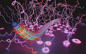
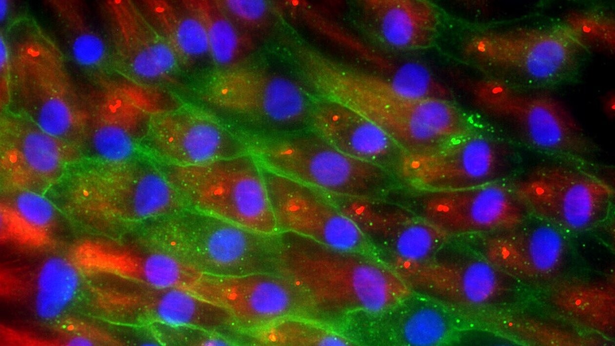
The malaria pathogen Plasmodium is transmitted from the mosquito’s salivary glands into the skin of the host. At this early stage, the single-celled parasite has a crescent shape. This unusual cell shape is responsible for the characteristic helical movements of the so-called sporozoites. They make it easier for the pathogen to curl around blood vessels or to get a grip in the surrounding tissue, as physicist Prof. Dr Ulrich Schwarz and malaria researcher Prof. Dr Friedrich Frischknecht demonstrated in earlier collaborative work. “Our new investigations show that malaria parasites move almost exclusively on right-handed helices in three-dimensional environments,” explains Prof. Schwarz, who heads the Physics of Complex Biosystems research group at the Institute for Theoretical Physics at Heidelberg University.
In experiments at the Center for Infectious Diseases of Heidelberg University Hospital, the scientists explored what biological function this right-handed motion could have. As a tissue substitute, they used synthetic hydrogels, which support the use of high-resolution imaging processes and a quantitative comparison with computer simulations of the cell movement. In the process, the researchers discovered that the parasites at the bottom of the hydrogel on the glass substrate behave differently than if applied to a glass slide directly from a fluid solution. In the first case, the parasites rotate clockwise on the glass; in the second, they rotate counterclockwise. Based on this, the researchers concluded that the right-handed motion is key to how the parasite penetrates different compartments.

“We suspect that this chirality developed during evolution to allow the pathogen to switch between the different tissue compartments in the host body quickly and always in the same way,” explains Friedrich Frischknecht, professor of integrative parasitology at the Medical Faculty Heidelberg of Heidelberg University and researcher at the Center for Integrative Infectious Diseases Research of Heidelberg University Hospital. The different movement patterns on conventional substrates in solution and coming from a three-dimensional hydrogel could explain why sporozoites were so poor at infecting liver cells in previous lab experiments. “Our results show that it makes a big difference if the pathogens are applied directly to the glass or if they first move through a tissue,” adds Dr Mirko Singer, a postdoc in Prof. Frischknecht’s group. The current findings on parasite movement could therefore help improve experimental assays and develop new approaches to infection prevention.
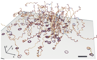
By combining high-resolution imaging and mathematical models, the researchers were also able to uncover the underlying molecular mechanism. Previous theoretical work had revealed how the parasite’s special crescent shape determines its motion. “Our computer simulations confirmed that only an asymmetry at the front end of the parasite could be responsible for the experimentally observed movement patterns,” states Leon Letterman, a doctoral candidate in the group led by Prof. Schwarz. Using super-resolution microscopy, the researchers identified a distinctive feature in the body plan of the parasite that results in an uneven force distribution along the body.
The research was funded by the German Research Foundation (DFG) and conducted in the framework of the Collaborative Research Centre “Integrative Analysis of Pathogen – Replication and Spread” based at the Medical Faculty Heidelberg of Heidelberg University. It was also part of the DFG-funded priority program “Physics of Parasitism”. Researchers from Johns Hopkins University in Baltimore (USA) also collaborated on the work. The results were published in the journal “Nature Physics”.
Original publication
L. Lettermann, M. Singer, S. Steinbrück, F. Ziebert, S. Kanatani, P. Sinnis, F. Frischknecht, U. S. Schwarz: Chirality of malaria parasites determines their motion patterns. Nature Physics (24 November 2025)